Looking at the surface of a membrane
What does the the membrane that wraps around our cells look like? We know that the membrane is choc-a-bloc full of proteins, but we can't see them directly because the level of detail is too small for our electron microscopes to look at, without destroying the cell.
Is there a way to re-construct the mosaic of proteins that normally stud a membrane? In "Molecular Anatomy of a Trafficking Organelle" Cell (2006) 127:831, Takamori and co-workers studied the synaptic vesicle - a little bubble wrapped up in membrane that carries neuro-transmitters from one synapse in the brain to another. 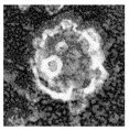 This is a synaptic vesicle as seen with electron microscopy. You can't go into any more detail. Instead, Takamori and co-workers determined the precise composition of all the proteins that float in the membrane of the synaptic vesicle. With this information, they built this delightful model of a synaptic vesicle:
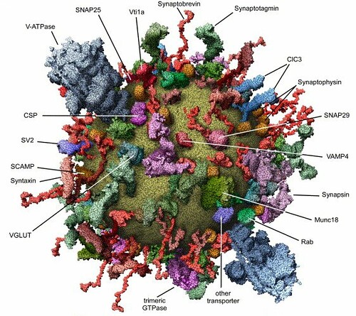
This image is somewhat reminiscent of the vibrant watercolors painted by David Goodsell, which were created using some deep intuitions about protein density and oodles of artistic license:![]()
![]()

1 comment:
Have you seen this short kinesin animation?
Post a Comment